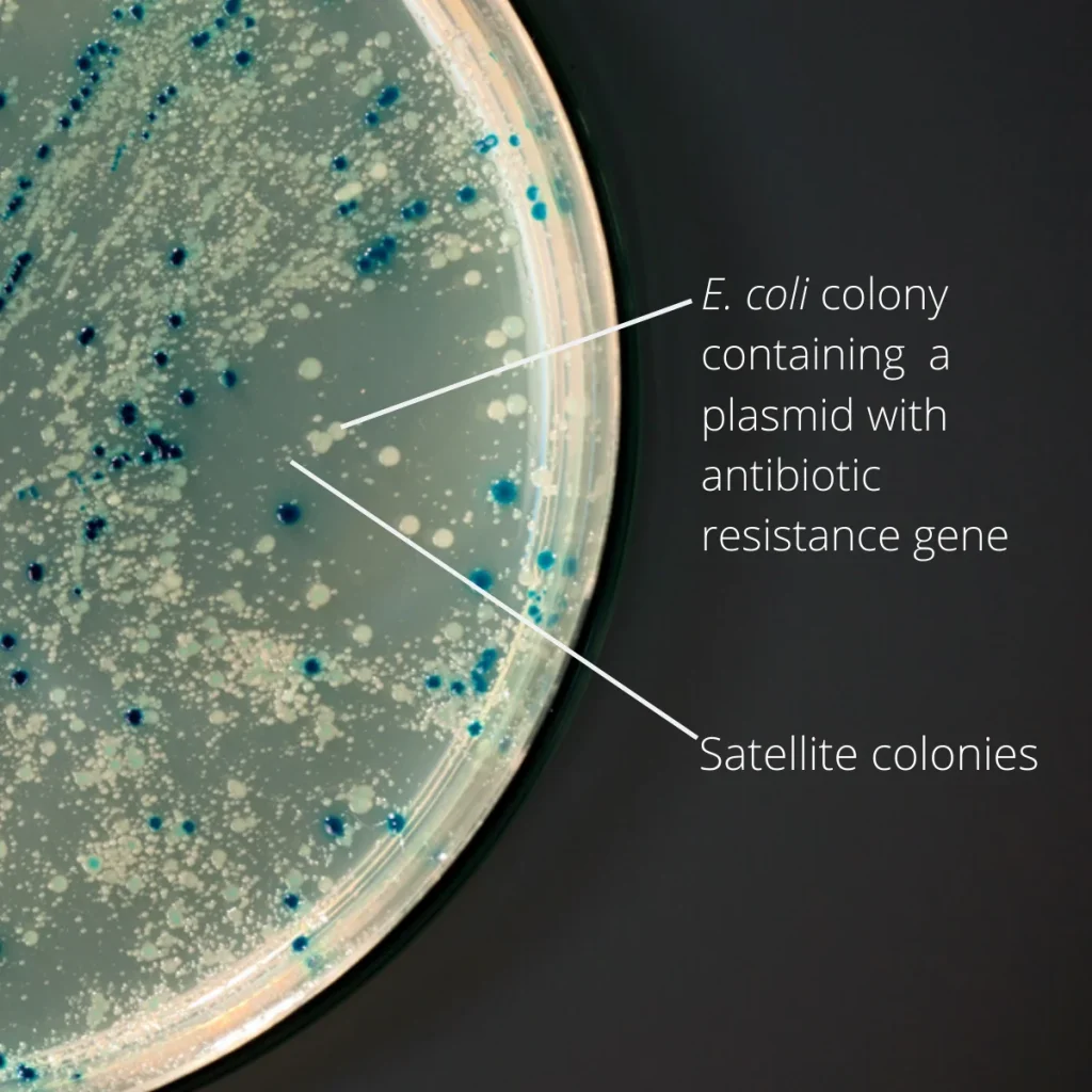
During microbiological examination, small colonies may sometimes appear around or close to larger microbial colonies on an agar plate. This pattern is known as the satellite phenomenon or satellitism.
Satellite colonies are usually smaller than the surrounding colonies and may depend on substances released or made available by nearby microorganisms for their growth. Recognizing this pattern is important because it can provide useful information about microbial interactions and help prevent incorrect interpretation of culture results.
In pharmaceutical microbiology laboratories, unusual colony patterns should be carefully documented and investigated rather than automatically classified as contamination or dismissed as insignificant.
What Are Satellite Colonies?
Satellite colonies are small microbial colonies that develop in the vicinity of larger colonies because the nearby microorganism provides growth factors or other conditions required for their development.
A well-known example occurs when Haemophilus influenzae grows around Staphylococcus aureus on blood agar.
H. influenzae requires two growth factors:
- Factor X: Haemin.
- Factor V: Nicotinamide adenine dinucleotide (NAD).
Although blood agar contains factor X, the availability of factor V may be limited. S. aureus can cause red blood cell lysis, making NAD available in the surrounding medium. This supports the growth of small H. influenzae colonies around the larger staphylococcal colonies.
This growth pattern is called the satellite phenomenon.
How to Identify Satellite Colonies on Agar Plates
Satellite colonies can be recognized through careful examination of their location, size and relationship with nearby microbial growth.
1. Observe the Colony Distribution
Look for small colonies growing close to or surrounding larger colonies.
In the classical satellitism pattern, smaller colonies are concentrated near the larger bacterial growth rather than distributed uniformly across the agar surface.
2. Compare Colony Size and Appearance
Compare the small colonies with the surrounding larger colonies.
Observe:
- Colony size and shape.
- Colour and pigmentation.
- Surface appearance.
- Elevation and margin.
- Distribution across the agar plate.
Differences in colony morphology may indicate the presence of more than one microbial population.
3. Examine the Growth Pattern
Check whether the smaller colonies are concentrated near a particular organism or streak.
In the classical H. influenzae example, satellite growth occurs near S. aureus on blood agar.
However, proximity alone does not confirm satellitism. Other growth patterns, mixed cultures and contamination may produce similar appearances.
4. Perform Further Identification
Colony morphology is only a preliminary observation. Further examination may include:
- Gram staining.
- Appropriate subculture.
- Biochemical identification.
- Validated microbial identification methods.
The identification approach should be selected according to the organism, sample type and laboratory procedure.
Satellite Colonies vs Other Small-Colony Growth
Not every small colony near a larger colony represents true satellitism.
| Observation | Possible explanation |
|---|---|
| Small colonies clustered around a larger colony | Possible satellite phenomenon |
| Small colonies distributed throughout the plate | Independent microbial growth or mixed culture |
| Small colonies appearing along a streak | Growth pattern related to inoculation or organism characteristics |
| Tiny colonies with delayed appearance | Slow-growing or nutritionally demanding microorganisms |
| Spreading growth around colonies | Possible swarming or other spreading growth behaviour |
The actual cause must be confirmed through appropriate microbiological investigation.
How to Handle Satellite Colonies in a Microbiology Laboratory
When a satellite-like growth pattern is observed, follow the approved laboratory procedure.
Step 1: Preserve the original observation
Do not immediately discard or disturb the plate. Record the unusual growth pattern and capture an image if permitted by the laboratory procedure.
Step 2: Document colony characteristics
Record relevant observations, including:
- Sample or plate identification.
- Culture medium used.
- Incubation conditions.
- Colony size, colour and appearance.
- Location of small colonies relative to larger colonies.
Step 3: Assess the significance
Review the sample source, test purpose and expected microbial profile. Determine whether the observed pattern could represent a known satellite phenomenon, mixed microbial growth or an unexpected contamination event.
Step 4: Perform appropriate identification
Where required, select representative colonies for further examination using approved methods. Small and large colonies should be evaluated separately when they appear to represent different microbial populations.
Step 5: Review and interpret the results
Compare the findings with the applicable test requirements, laboratory specifications and historical observations. Do not identify an organism based on colony appearance alone.
Step 6: Document the investigation
If the growth is unexpected or affects the interpretation of the test, document the assessment and follow the laboratory’s deviation or investigation procedure.
Importance of Satellite Colony Identification in Pharmaceutical Microbiology
Recognizing unusual colony patterns is important in pharmaceutical microbiology because microbial growth may be detected during:
- Environmental monitoring.
- Microbial limit testing.
- Sterility testing.
- Water microbiological testing.
- Culture media performance assessments.
For example, if small colonies appear near larger colonies on an environmental monitoring plate, the laboratory should determine whether they represent separate organisms or a genuine satellite growth pattern.
Incorrect interpretation may result in incomplete microbial identification or an inaccurate understanding of the contamination profile.
Therefore, unusual colony morphology should be evaluated using appropriate identification techniques and documented according to the approved SOP.
Common Mistakes to Avoid
- Assuming every small colony is a satellite colony.
- Identifying microorganisms only through colony morphology.
- Ignoring small colonies growing near larger colonies.
- Selecting only the larger colonies for identification when multiple morphologies are present.
- Discarding the original plate before documenting unusual growth.
- Performing additional tests without following approved laboratory procedures.
- Failing to investigate unexpected microbial growth.
Frequently Asked Questions (FAQs)
1. What causes satellite colonies on agar plates?
Satellite colonies develop when microorganisms depend on growth factors or other substances made available by nearby microbial growth.
2. Which organism commonly demonstrates the satellite phenomenon?
Haemophilus influenzae is a classical example. It can form small satellite colonies around Staphylococcus aureus on blood agar because NAD (factor V) becomes available through red blood cell lysis.
3. Are satellite colonies always a sign of contamination?
No. Satellitism is a recognized microbial growth phenomenon. Its significance depends on the organism, culture medium and sample source.
4. Can satellite colonies be identified by appearance alone?
No. Colony morphology provides preliminary information, but reliable identification requires appropriate microbiological tests.
5. Should small satellite-like colonies be included in microbial counts?
Where the test requires enumeration, all countable colonies should be evaluated according to the applicable method. The laboratory should not exclude small colonies simply because they appear near larger colonies.
Conclusion
Satellite colonies represent an interesting microbial growth phenomenon in which one microorganism supports the growth of another by making essential growth factors available.
Recognizing this pattern helps microbiologists distinguish potential satellitism from mixed microbial growth and other unusual colony appearances. In pharmaceutical laboratories, careful observation, appropriate identification and complete documentation are essential for reliable microbiological results.
References
- American Society for Microbiology (ASM). Microbiology Laboratory Tips and Tricks: An Organism Isolation and Identification Survival Guide. Read article
- Centers for Disease Control and Prevention (CDC). Public Health Image Library: Haemophilus influenzae Satellite Colonies Around Staphylococcus aureus. View image and description
- UK Health Security Agency / Royal College of Pathologists. UK Standards for Microbiology Investigations: Identification of Haemophilus Species and the HACEK Group of Organisms. Read guideline
- NCBI Bookshelf. Haemophilus Species. Read microbiology reference