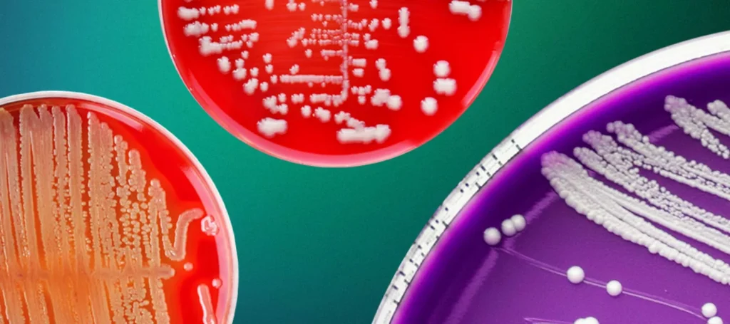
During microbiological testing, bacterial colonies may develop different colours as they grow on agar plates. Some colonies appear white or cream initially and later develop yellow, red, green or other pigmentation. In certain cases, the surrounding agar also changes colour.
These changes may occur due to bacterial pigment production, metabolic activity, enzyme reactions or interactions with components of the culture medium. Observing colony colour is an important part of microbiological examination because it provides useful preliminary information about microbial characteristics.
In pharmaceutical microbiology laboratories, however, colony colour should not be used as the only basis for microbial identification. Further testing may be required to confirm the organism.
What Causes Bacterial Colonies to Change Colour?
Bacterial colony colour is influenced by the organism’s characteristics, culture medium and incubation conditions. The following are some common reasons for colour development during incubation.
1. Production of Bacterial Pigments
Some bacteria naturally produce coloured substances called pigments during their growth.
Pigment production may become more noticeable as the bacterial population increases or as the organism produces more pigment over time.
Examples include:
- Staphylococcus aureus – commonly produces golden-yellow colonies.
- Micrococcus luteus – produces yellow-pigmented colonies.
- Serratia marcescens – can produce a red pigment called prodigiosin under suitable conditions.
- Pseudomonas aeruginosa – may produce blue-green pigments, including pyocyanin.
The appearance and intensity of pigmentation can vary depending on the culture medium and incubation conditions.
2. Changes in pH Due to Bacterial Metabolism
Bacteria utilize nutrients present in culture media and produce metabolic end products.
When carbohydrates are fermented, acidic products may be formed, lowering the pH of the medium. Some culture media contain pH indicators that respond to these changes by producing a visible colour reaction.
For example:
- On MacConkey agar, lactose-fermenting organisms produce pink to red colonies due to acid production and the neutral red indicator.
- On certain differential media, changes in pH may produce a visible colour change around bacterial growth.
Therefore, a colour change may reflect bacterial metabolic activity rather than pigment production.
3. Enzyme Activity in Chromogenic Media
Chromogenic culture media contain specific substrates that produce coloured compounds when acted upon by microbial enzymes.
During incubation, bacterial enzymes break down these substrates and release coloured products.
For example, certain chromogenic media use substrates that detect enzymes such as beta-glucosidase or beta-galactosidase.
The resulting colony colour helps differentiate microorganisms based on their enzymatic activity.
However, chromogenic reactions are not always species-specific. Additional identification may be necessary.
4. Influence of Incubation Temperature
Temperature can influence bacterial growth and pigment production.
Some microorganisms produce visible pigments only within particular temperature ranges. Changes in incubation temperature may affect the intensity or appearance of colony pigmentation.
For example, Serratia marcescens may produce a prominent red pigment at lower temperatures, while pigment production may be reduced at higher temperatures.
Therefore, differences in incubation conditions can lead to variations in colony appearance.
5. Changes in the Surrounding Culture Medium
Not all colour changes occur within the bacterial colony itself. Some microorganisms release substances that react with or diffuse into the surrounding agar.
Examples include:
- Pigments that diffuse through the medium.
- Acid production that changes the colour of pH indicators.
- Hydrogen sulphide production that causes blackening in suitable media containing appropriate indicator components.
- Haemolytic activity that produces visible changes around colonies on blood agar.
These reactions provide additional information during microbiological examination.
Examples of Bacterial Colony Colour Changes
| Bacterium | Common colour or appearance | Possible explanation |
|---|---|---|
| Staphylococcus aureus | Golden-yellow | Pigment production |
| Micrococcus luteus | Yellow | Pigment production |
| Serratia marcescens | Red | Prodigiosin production under suitable conditions |
| Pseudomonas aeruginosa | Blue-green | Pigment production |
| Escherichia coli | Pink to red on MacConkey agar | Lactose fermentation |
| Vibrio cholerae | Yellow on TCBS agar | Sucrose fermentation |
These are typical examples, not universal identification criteria. Colony appearance can vary with the medium, strain and incubation conditions.
How to Examine Colour Changes During Incubation
When an unusual colour change is observed, microbiologists should examine the plate systematically.
Step 1: Observe the initial appearance
Check the original colour of the agar and record the appearance of the colonies when first examined.
Step 2: Compare colony and medium colour
Determine whether the colour is limited to the colony or has spread into the surrounding agar.
Step 3: Record changes in colony morphology
Observe pigmentation along with colony size, shape, margin, elevation and surface characteristics.
Step 4: Review incubation conditions
Check the incubation temperature, duration and other conditions against the approved test procedure.
Step 5: Perform further identification
If the colour change is unusual or relevant to the investigation, use appropriate identification methods such as Gram staining, biochemical tests or validated automated identification systems.
Step 6: Document the observations
Record the findings according to the laboratory SOP and applicable documentation requirements.
What Should Be Done If a Colony Changes Colour Unexpectedly?
Unexpected colony pigmentation should be assessed rather than immediately considered contamination or a confirmed identification result.
The laboratory should:
- Preserve the original observation and plate identification details.
- Check whether the medium is suitable and within its approved use period.
- Review the incubation conditions and preparation records.
- Compare the colony appearance with previous observations, where available.
- Perform further identification when required.
- Initiate an investigation if the result is unexpected or affects the test outcome.
In pharmaceutical microbiology, the significance of unusual growth should be assessed according to the sample type, test purpose and approved investigation procedure.
Common Mistakes to Avoid
- Identifying bacteria based only on colony colour.
- Assuming every colour change is caused by pigment production.
- Ignoring changes in the surrounding agar.
- Comparing colonies grown on different media without considering medium composition.
- Overlooking incubation temperature and duration.
- Recording unusual pigmentation without documenting the medium and incubation conditions.
- Discarding unexpected growth without appropriate evaluation.
GMP Importance of Observing Colony Colour
Colony morphology and pigmentation are useful observations during microbiological examination in pharmaceutical laboratories.
They can help identify unusual microbial growth patterns during environmental monitoring, microbial limit testing and other microbiological investigations.
However, colony colour alone cannot establish microbial identity or determine the source of contamination.
A systematic approach involving observation, documentation and appropriate microbial identification supports reliable laboratory results and effective contamination investigations.
https://pharmavk.com/satellite-colonies-on-agar-plates/
Frequently Asked Questions (FAQs)
1. Why do bacterial colonies become more colourful during incubation?
Some bacteria produce pigments as they grow. As pigment accumulates, the colonies may become more visibly coloured.
2. Does colony colour depend on the culture medium?
Yes. Nutrient composition, pH indicators and chromogenic substrates can influence colony colour and the appearance of the surrounding agar.
3. Why does Serratia marcescens produce red colonies?
It can produce a red pigment called prodigiosin. Pigment production is influenced by environmental conditions, including temperature and culture conditions.
4. Can bacterial pigments spread into the agar?
Yes. Some pigments are water-soluble and diffuse into the surrounding medium, while others remain largely within the bacterial colonies.
5. Can bacterial species be identified by colony colour alone?
No. Colony colour is a preliminary characteristic. Reliable identification requires additional microbiological or validated identification methods.
Conclusion
Bacterial colonies may change colour during incubation because of pigment production, metabolic activity, enzyme reactions and interactions with culture media. These changes provide useful preliminary information about microbial growth and characteristics.
For pharmaceutical microbiology laboratories, careful observation of colony pigmentation, documentation of unusual findings and appropriate microbial identification are important for reliable test interpretation.